West Nile Virus (WNV)

Causative Agents

West Nile virus (WNV) is a Flavivirus, belonging to the Flaviviridae family that was first isolated in 1937 in “West Nile” province of Uganda/Africa and was named after this region. WNV virions are approximately 50 nm in diameter and are composed of a single-stranded RNA genome of length between 11kb and 12 kb. [1]

Epidemiology

WNV was originally isolated in the West Nile region of Uganda in 1937. It was then identified in Congo, Sudan, India, Egypt and Israel. Later on, infection spread outside Africa and was eventually found to be widespread in Africa, West Asia, Middle East, Southern Europe, Australia and US. Several WNV outbreaks has occurred in Israel (1957, 2000), US (1999, 2003-2007), Romania (1996-1997) and Canada (2002-3003). Recent WNV outbreak in Greece in 2010 with more than 10 deaths has been reported. WNV infection has been identified to be the cause of 3 deaths between August and September of 2010 in Turkey. Although WNV was first emerged as a distinct virus, currently it is classified into two lineages where lineage 1 is associated with clinical human encephalitis and lineage 2 with several outbreaks in horses. [2], [3]

Modes of Transmission

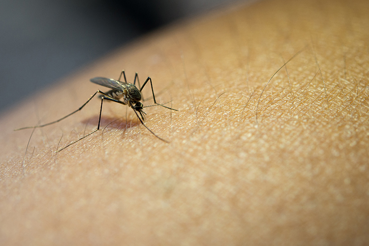
Most of the WNV infections are asymptomatic without any symptoms in people. Second type WNV infections result in mild fever (West Nile fever) with recovery from the illness ,but however, third type of WNV infections with the virus crossing the blood-brain barrier cause severe outcomes such as inflammation of the brain (encephalitis) and the tissue surrounding the brain and spinal cord (meningitis) that all resulting in long-term effects like memory loss, depression etc.. The common route of WNV transmission is shown to be the mosquitos as vectors carrying the virus across vertebrate hosts. Mosquitos receive the virus upon feeding on infected birds, and then virus is transmitted to other hosts via mosquito bites. Humans, horses and most other mammals are widely accepted as incidental hosts as they don’t transmit virus to others. However, with increasing research after WNV outbreaks, new WNV transmission routes has been shown to be transmitted via blood transfusion, organ transplantation, intrauterine exposure and breast feeding. [3]

Diagnosis

Differential diagnostic tests, such as plaque-reduction neutralization test (PRNT), ELISA, hemagglutinin inhibition test, indirect immunofluorescence test, are applied for identification of WNV. ELISA is one of the frequently used methods involving detection of antibodies against WNV antigens affixed to a surface in samples of serum or cerebrospinal fluid. It is easy and with high sensitivity. But not able to presume quantitative results and also may give false-positive results for immune-compromised patients. The recently applied technique, use of real-time PCR technology may complete the need of quantification in order to follow the clearance of the virus and thus evaluate the efficacy of therapeutic interventions. [4]

References

1) Brinton MA:  The molecular biology of West Nile Virus:  a new invader of the western hemisphere. Ann Rev Microbiol 2002;56:371-402

2) 2008 Final West Nile Activity Map, Centers for Disease Control and Prevention

3) Hayes EB, Komar N, Nasci RS, Montgomery SP, O’Leary DR, Campbell GL (2005). “Epidemiology and transmission dynamics of West Nile virus disease”. Emerging Infect. Dis. 11 (8): 1167–73.

4) Busch MP, Tobler LH, Saldanha J, et al:  Analytical and clinical sensitivity of West Nile virus RNA screening and supplemental assays available in 2003. Transfusion 2005;5(4):492-499

5) Epstein JS:  Insights on donor screening for West Nile virus. Transfusion 2005;45(4):460-462

6) New York City Department of Health. West Nile surveillance and control: an update for healthcare providers in New York City. City Health Information. June 2001;20(2)

Related Kits