Meningitis, defined as inflammation of the membranes surrounding the brain and spinal cord, is a disease that requires immediate medical attention. Fever, headache, and vomiting are the main symptoms of meningitis; however, these symptoms can also be detected in some other diseases. Therefore, in the presence of such symptoms, it is very important to apply to a health institution quickly.
Meningitis
Viruses are the most common cause of meningitis. However, bacteria, fungi, or parasites can also cause meningitis. Meningitis caused by viruses is the most common type and is less severe than others. The enterovirus family, which is generally associated with the digestive system, can cause meningitis with viruses that cause diseases such as chickenpox, measles, mumps, and herpes. Bacterial meningitis, although relatively rare, can have very serious consequences if left untreated. In addition to these, bacteria such as Streptococcus pneumoniae, Neisseria mengitidis, Haemophilus influenza type b, Escherichia coli, and group B streptococci are among the causes of meningitis.
Epidemiology
Although meningitis can be seen at any age, the risk is quite high in newborns and children younger than 1-year-old. Also, the incidence of meningitis is increasing in people over the age of 60. Childhood and later vaccinations protect against most disease-causing infections; yet, it is very important for parents to consider the symptoms of the disease, start the treatment without losing time, and prevent the disease from causing harm.
According to the WHO, bacterial meningitis is of particular concern. About 1 in 10 people who get this type of meningitis die and 1 in 5 people have serious complications; that is, these bacteria were responsible for more than 50% of the 250,000 deaths from all-cause meningitis in 2019. They also cause other serious diseases such as sepsis and pneumonia.
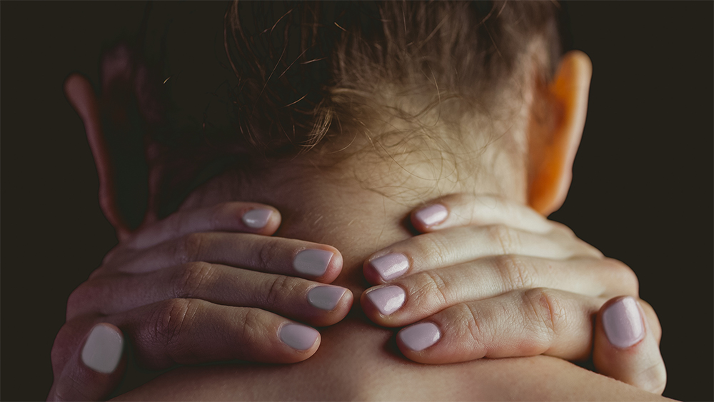
Symptoms
Child and adult symptoms are as follows;
- Fever of 38 degrees or higher
- Severe headache
- Stiff neck
- Avoid looking at bright light
- Drowsiness and unresponsiveness
- Clouding of consciousness
- Cold hands and feet
- Shake
- Joint and muscle pains
However, babies might show symptoms a little differently;
- Rapid breathing
- Eating less
- Groaning or high-pitched crying
- A pale and blotchy appearance
- Excessive tension and swelling of the fontanelle on the head
- Rigidity
Transmission
Viruses or bacteria enter the body through the nose, throat, or ear and reach the brain, causing an infection. The route of transmission is listed below:
- Coughing
- Sneezing
- Kissing
- Using communal utensils, plates, and cutlery.
Since the immune system is not developed in children, meningitis is one of the most dangerous diseases. In addition, people with damaged or removed spleens and people with health problems such as long-term illness or immune system disorders such as AIDS or cancer patients, are also at risk.
Because some germs that cause meningitis can spread easily, outbreaks can occur in densely populated areas where people live close together. The possibility of spreading the agent is high in places such as dormitories and barracks. In addition, it is recommended that adults who travel to regions such as Africa and India and who visit Hajj should have meningitis vaccination again.
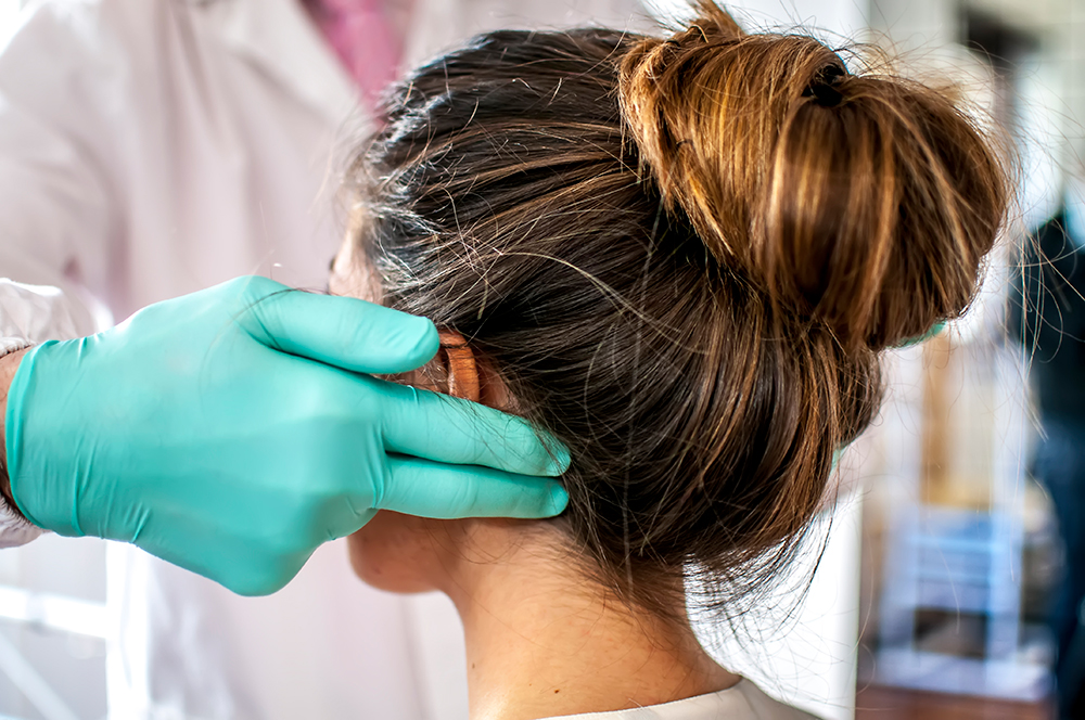
Diagnosis
Infectious meningitis can be caused by viral, bacterial, or fungal pathogens. Despite widely available treatments, many types of infectious meningitis are still associated with significant morbidity and mortality. A delay in diagnosis contributes to poor outcomes.
For the diagnosis, a physical examination is first performed by the doctor. Neck stiffness, flexibility of the joints, and skin rash are checked. Lumbar puncture procedure may be performed from the waist, cerebrospinal fluid is taken between the lumbar vertebrae with the help of a needle, and the presence of bacteria and viruses is investigated. Computed tomography is used to check for any anatomical changes in the brain. But above all, Real-Time PCR is a better option than others in terms of speed and sensitivity.

Treatment
Bacterial meningitis can be drug-treated when early onset of the disease. Also, there are some vaccines available for different meningitis pathogens.